|
|
|
|
|
|||
| 1 |
PREGNANCY TEST Scanner carried on the arm eases the task of diagnosing pregnancy. Because the complete scanner including the screen can be carried on the arm when carrying out pregnancy examinations, the testing of the sows becomes a lot more accurate and also more fun. It is Lisa Nyberg, who is self-employed within the insemination service, who says this. Lisa pregnancy tested almost 25 000 sows during 1999. The scanner shows the foetus in the uterus, and it is possible to "freeze" the image for a closer examination. |
||
| Lisa has previously worked with several different pregnancy-testers, a machine that emits a sound, Pregton, and others, like Doppler, which gives out a sound via headphones. Furthermore there is Ultra Scan, a scanner that is carried in a strap around the neck/shoulder and is supplied with a screen. - For me Tringa is the best scanner to test the pregnancy. It is easy to work with and is a lot more flexible than the others, says Lisa. | |||
| The foetus can be seen on the screen. | |||
| "For me it is important to be able to see whether the animal is pregnant or not", says Lisa. "When I perform a test with the scanner, a section of the abdomen of the animal and what is in it, is shown. If the sow is not pregnant a completely empty uterus with "black strings" is shown on the screen. If the sow is in heat, the picture of the uterus is muddled. Also if the sow is not well. It looks like a "snow storm" in the black ducts." "If needed, the picture on the screen can be "frozen" so that I am able to study the image over a longer period.", Lisa explains. In the case of pregnancy, liquid is pictured in the uterus. A Pregton that emits a sound in the case of liquid may also give out a sound in the case of cysts or if the animal is in heat. If the probe is placed to far back it can also sound to the liquid in the urine bladder. In the headphones of a Doppler pulsation can be heard, but it is not quite as "buzzing" from a uterus that contains cysts. The best way to learn to interpret the scanner image is to watch non-pregnant animals repeatedly to see how the uterus changes. Another possibly is, during slaughter, to carry out an autopsy of a uterus that has caused problems, and then find an answer to the scanner image. | |||
|
|
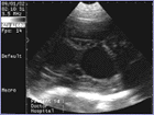
Picture: Screen image showing a 26-day-old foetus. The black "holes" are foetus sacks. |
||
| The screen on the lower arm | |||
|
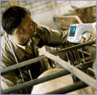
The scanner to test the pregnancy, the 50S Tringa, used by Lisa Nyberg, is fastened on the left lower arm. The scanner only weighs 800 g. A glove without fingers is first put on the hand and on to this glove the 50S Tringa is fastened with Velcro so that it is attached properly. Then Lisa pulls the cable from the scanner to the probe, first through the left arm of the overall and behind her back and then through the right arm. This way there is no cable for the sows to bite into. The battery for the scanner is fastened at the waist, for example to the belt. Even these wires follow the arm to the scanner. The battery has an approximate running time of four hours. With the scanner/screen on the arm it is much easier to see the image. The person can also have a bottle of gel in the same hand that the scanner is attached to. The gel is applied on the probe to get good contact with the skin. Even if the sow is restless and moves backwards and forwards in the pen or is loose, the person doing the scanning can position the probe correctly against the abdomen and at the same time position the arm, so it is possible to see the image on the screen. "This scanner is definitely simpler and easier to work with than a scanner that dangles around the waist, which may make the reading of the image more difficult. At the same time a scanner that hangs by a strap around the neck will take up space between the person and the pen. It can then be difficult to hold the probe in place and see the image on the screen at the same time. There is also a clear difference in the weight one is forced to carry around during the whole duration of the examination." |
|||
 |
Picture: Both hands are free to hold the probe and the bottle of gel. |
||
|
An average of 30 seconds |
|||
|
Because Lisa works among many different livestock, she has a separate glove (the one that holds the screen) for different livestock and a few spare gloves that she washes between each use. Then there is less risk of infection spreading between livestock. The scanner is wiped clean and disinfected between each use "Without exaggerating, it takes on average no more than 20 - 30 seconds to test a sow on pregnancy", tells Lisa. The average is shorter the more sows that are tested. "The best time for pregnancy testing is 4 - 5 weeks into the pregnancy. Then I am able to quickly see the foetus sacks on a pregnant animal. Already from 8 weeks or more the skeleton of the foetus can be clearly seen on the screen. It may be a little difficult to read 6 -7 weeks into the pregnancy. No skeleton can yet be seen and the foetus sacks looks quite small. Pregnancy testing among bigger livestock is a necessity. Empty days and re-insemination is expensive. One empty day can be valued at SKr 50. A scanner can also be used to examine sows as well as pigs to see if more pigs are left in the uterus.
|
|||
|
The purchase price must be seen in relation to many factors |
|||
|
Different aspects in the use of the scanner must decide if this is expensive or less expensive. The purchase price must be seen in relation to an improved working environment where the scanner is used, simpler and faster work with improved efficiency, and less empty and lost days among the livestock, and when used in connection with the farrows. One empty day is estimated to approx. SKr 50. If you have approximately 500 sows among your livestock that are tested 2.25 times a year, you are testing approx. 1125 sows. That works out at about Skr 17 per animal with the high purchase price and written off over a period of three years. Interest is not included! If the purchase price is spread out over the number of empty days it will be 1180 days. The costs saved when using the Tringa for other examinations (see below) are not included. In reality you end up with a combination of all the "ingredients". The scanner can also be used at the farrowing to see if any pigs are left in any part of the uterus. This can be the first alternative before examination. Manual examination always carries the risk of infection, which often involves treatment with antibiotics that costs time and money. Apart from pigs, the scanner can also be used for horses, sheep and even cattle. On the bigger animals like horses and cattle, rectal examination is used because this gives the shortest distance between the probe and the uterus, we are told by Kruuse, the representative of the Pie Medical products in Sweden. The scanner can be equipped with different probes that have different frequencies. According to Kruuse a higher frequency gives a better and clearer screen image and a higher resolution. Lower frequencies reach deeper into the tissue. Normally the scanners are equipped with a probe with frequencies that reach deeper into the tissue. This article
has been printed with the permission of: |
|||
| 2 | DEMONSTRATING THE USE OF THE TRINGA | ||
|
Lisa Nyberg, who has a long experience in the use of the Tringa, gave a demonstration for Katarina Persson, at Tranums pool of sows, west of Lidköping. First Lisa performed a pregnancy test of the sows and pointed out the different features of the scanner until Katarina learned how to use it. In addition to the testing of sows at 4 weeks into pregnancy, the sows of which the pregnancy is uncertain will be tested again at 11 weeks, the week before the sows are to be transported to the "satellite". In this way the annoyance of sending empty sows to the "satellite" is avoided. Tranum's pool of sows is one of the five in Sweden that has purchased a Tringa. There are already more than 300 sold abroad. |
|||
|
|
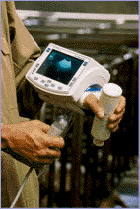
Picture: By keeping the arm bent it is easy to see if the sow is pregnant or not on the screen. | ||